Equine medicine through the ages
Veterinary medicine has seen rapid developments in recent decades. In James Herriot’s time, vets were all-round general practitioners who treated any disease in pets, farm animals and exotic beasts. With the continuous advancements in veterinary medicine, it is hardly possible today for one vet to cover all possible specialty areas in all species lege artis, i.e. in line with current science. Veterinary medicine has become more specialised over the decades, just like human medicine. At the same time, the profession has undergone a gender change: veterinary medicine was once a traditionally male profession, but today it is mostly women who find their way into the demanding study of veterinary medicine.
The first point of contact for horse owners used to be the general practitioners mentioned above; that is, vets in large-animal or mixed practice. These days, however, this role is more often taken on by practitioners with an equine focus and with additional training in equine medicine and surgery.
These veterinarians offer solid, basic medical care for horses in all fields of equine practice.
Various areas have evolved as a further level of specialisation with internationally recognised special training and qualifications. This training represents the highest level of clinical qualification in veterinary medicine and is goverened by guidelines from European or American ‘Colleges’. Alongside international networking and lively knowledge exchange between specialists, the Colleges are represent state-of-the-art veterinary medicine and also provide quality assurance and promote ongoing medical advances. The specialties regulated by the Colleges include surgery, internal medicine, emergency and intensive care medicine, and sports and rehabilitation medicine. There are also cross-species specialist areas relevant to equine practice. These include, for example, cardiology, ophthalmology, reproductive medicine, anaesthesia and pain therapy, dermatology, neurology, oncology, diagnostic imaging, laboratory medicine and animal nutrition. Ideally, large modern veterinary clinics now work on an interdisciplinary basis, with a variety of these specialists caring for the patients together. Veterinary medicine at these clinics is largely teamwork.
Because horses are large animals, a top-class infrastructure and modern instruments and equipment are needed to treat the patients appropriately, in addition to the staff requirements. Since today horses are increasingly not treated in the stables, but are brought to a clinic for diagnosis and treatment, special attention is also given to the safe transport of the horse to the hospital or clinic facility. Primarily in emergency situations, such as cases of colic or fractures, owners and vets today frequently rely on trained professionals who can provide specially equipped transport vehicles. These include various suspension systems for rescue and transport, but also mattresses so horses can be transported even when lying down.
“Why had I entered this profession? I could have gone in for something easier and gentler – like coalmining or lumberjacking. If you decide to become a veterinary surgeon you will never grow rich but you will have a life of endless interest and variety.”
James Herriot (1916–1995), British veterinarian and author

Veterinary care in a horse’s life
Foals, which nowadays can also be bred by in vitro fertilisation and embryo transfer, are often already monitored during the pregnancy of the mare. They can suffer from complex problems during and immediately after birth, or in the first weeks and months of life. Just as in human medicine, foals can be fully cared for in neonatal intensive care units by emergency and intensive care specialists and internal medicine experts, and they can even be fed intravenously if required. Furthermore, foals may need to undergo surgery at an early age, for example for ruptured bladders, umbilical infections, fractures or colic. These small, vulnerable patients need to be comprehensively assessed, stabilised, and prepared appropriately for surgery; they need a tailored general anaesthesia, an experienced surgeon and meticulous, intensive postoperative care from intensive care specialists.
Throughout a horse’s life, veterinarians are an essential part of the team involved to maintain health and prevent disease, with prophylactic check-ups provided on a regular basis. In addition, horses are frequently injured or develop diseases that require urgent veterinary attention in order to save or preserve the horses life. The most common preventative measures include nutritional advice, vaccinations, deworming, dental treatment and castration. These treatments are usually conducted by equine veterinarians or general veterinary practitioners in the stables environment. However, it is becoming more and more common to bring older stallions to a clinic for castration, so that the procedure can be conducted by a specialised surgeon and an anaesthetist in a sterile operating theatre. This reduces the risk of complications and accelerates post-operative recovery.
Any horse may suffer from injuries or diseases of the musculoskeletal system, dental diseases, gastrointestinal, respiratory and cardiovascular diseases, nervous system disorders, hormonal and metabolic disorders, urinary complaints, infections and skin diseases.
Clinical examinations are an important part of equine medicine, and remain the basis for diagnosis and treatment. The use of a variety of new diagnostic procedures enables the clinician to obtain a faster and more accurate diagnosis, helps in selecting the appropriate treatment and provides the clinician with more information when determining the prognosis.

Today, transport and rescue services for large animals provide indispensable assistance during emergency transport by road and air. (Pictures: Large Animal Rescue Service Switzerland/ Liechtenstein®) Foal medicine is often complex and requires extensive knowledge of internal medicine and intensive care medicine. (Picture: A veterinarian and a prospective veterinarian of the Equine Hospital examining a sick foal.)


Diagnostic and therapeutic methods
In recent years, disease diagnosis has been significantly improved by the use of advanced X-ray and ultrasound equipment, as well as by scintigraphy, computer tomography (CT)
and magnetic resonance imaging (MRI). X-rays have long been standard for diagnosing many common diseases, such as lung diseases, orthopaedic diseases or diseases of the head. Modern, high-resolution ultrasound devices have become more affordable for equine practitioners in recent years and are therefore widely used now. They help diagnosing soft tissue problems, such as tendon disorders, heart disease or diseases of the abdominal organs. For diseases of the liver, kidneys, spleen, lungs or deeper lymph nodes, ultrasound-assisted tissue samples can be obtained from the various organs, allowing a definitive diagnosis to be made and a prognosis established.
In addition, advanced imaging techniques such as MRI, CT and scintigraphy play a central role in diagnosing orthopaedic and neurological conditions, head and dental diseases as well as diseases in many other organs. Generally, these procedures are only available at larger clinics. The sheer variety of imaging techniques and special procedures for each intervention means that extensive training and experience is required for evaluating and interpreting the test results correctly. These advanced methods in particular should only be used by qualified specialists in order to avoid misinterpretations and incorrect or missed diagnoses.
Imaging techniques, however, are not just diagnostic, but are increasingly used in treating various diseases. In orthopaedics, modern implants are inserted using computer-assisted surgical navigation or CT guidance. This means that screws for stabilising broken bones and hairline fractures can be placed with millimetric precision. Bone cysts, too, can be successfully treated in difficult-to-access parts of the anatomy in this way. The boundaries of what is possible in orthopaedic surgery have been pushed to a much higher level as a result.
Fungal diseases, which damage the arteries in the horse’s guttural pouches and can cause life-threatening bleeding, can be precisely treated with the help of a C-arm, a special device using X-ray technology, and fluoroscopic guidance. In this procedure, a catheter is used to insert coils or vascular plugs into the affected blood vessels; this closes them and stops the bleeding. Finally, certain heart rhythm disorders or cardiac malformations can also be specifically treated using cardiac catheters with X-ray or ultrasound guidance.
More and more interventions are becoming minimally invasive, using keyhole techniques in order to reduce postoperative pain and avoid complications. These minimally invasive procedures principally include arthroscopy (to visualise joints) and laparoscopy (to visualise the abdominal cavity); procedures such as thoracoscopy (visualisation of the thoracic cavity) are used less often.
Today, a large number of joint diseases can be successfully treated using arthroscopy. Various laparoscopic techniques are used to diagnose gastrointestinal diseases or diseases of other internal organs, or to permanently remedy recurrent types of colic such as the displacement of the intestines into the nephrosplenic space or the epiploic foramen, a gap between different abdominal cavity areas.
Minimally invasive procedures also include endoscopic lithotripsy, that is, the destruction of urinary stones with high-energy ultrasound.
In respiratory surgery, diode lasers or electocautery devices enable vets to perform minimally invasive interventions on a standing horse using an endoscope. This technique, alongside operations like laryngoplasties (to treat laryngeal paralysis) and tie-forwards (to treat dorsal displacement of the soft palate), are the mainstay of respiratory surgery.
In recent years, equine dentistry has developed into an
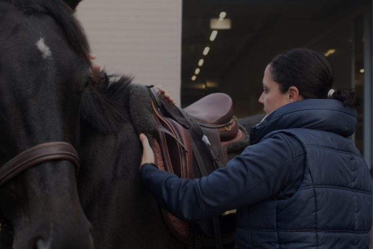
independent field of expertise. (Picture: Veterinarian examining the mouth and teeth of a horse)The health of the horse’s back is central to the performance
of a sport horse. (Picture: Veterinarian examining the saddle fit of a horse)


While viewing internal, hard-to-access hollow organs was not previously possible in living animals, vets today can reach for a number of high-resolution rigid or flexible endoscopes of varying thickness and length and use them for diagnostic and therapeutic purposes. In this way, the airways including the guttural pouches, the oral cavity, the pharynx, the rectum, the urinary tract including the bladder, and even the outer ear canal can all be examined with relative ease. Flexible video endoscopes up to 320 cm in length are used in gastroscopies on adult horses. These endoscopes can be used to reach as far as the duodenum and they are also designed for taking tissue samples from the stomach and bowel.
As certain diseases of the upper respiratory tract are dynamic, and emerge particularly as the result of exercise, affected horses are today examined not just at rest but also when moving. This can be done either as a field test with a rider, using a mobile, portable endoscopy unit, or on a high-speed treadmill using a stationary endoscopy system. Functionally unclear findings can also be dynamically investigated with a spirometry system, which quantitatively assesses lung function to establish the degree of airway obstruction. Once upon a time, all this would have been unimaginable.
Horses often suffer from heart murmurs and heart rhythm disorders. While some of these can be classified as “normal”, or “pathological but relatively harmless”, others result in a significant increase in the risk of an adverse event. Such cases must be identified and assessed at an early stage, and many non-invasive examination options are available for this purpose. Blood pressure measurements, cardiac ultrasound examinations and electrocardiography (ECG, including exercising ECGs and 24-hour resting ECGs) are part of routine diagnostics, although correctly conducting and assessing the findings requires special training and a great deal of experience, which is only available at a few larger clinics in Europe. Exercise tests in cardiac patients can be conducted, just like airway tests, either in the field with a rider or on the lunge, or on a high-speed treadmill under controlled conditions.
Previously, horses with atrial fibrillation were treated with medication that had significant adverse effects. Today, transvenous electrical cardioversion is available at selected clinics for these patients. This treatment virtually restarts the heart’s electrical conduction system using an electric shock. Even implanting a pacemaker in a horse with a cardiac arrhythmia can now be done without difficulty. Treating certain heart rhythm disorders with electrical mapping, followed by catheter ablation – the destruction of the pathological tissue using a catheter – is currently in the early stages of development.

Today, specialists are also available with state-of-the-art instruments and equipment for high-quality diagnosis and treatment of eye diseases and injuries. Patients with equine recurrent uveitis (moon blindness) can be treated by vitrectomy, in which the vitreous body is surgically removed from the eye, or with implants that release the immunosuppressant cyclosporine. A few centres also offer cataract operations by implanting artificial lenses, the surgical treatment of glaucoma and the stabilisation of corneal injuries using UV-radiation. In addition, they also use different laser devices for treating various eye diseases and state-of-the-art imaging techniques such as magnetic resonance imaging, high-resolution ultrasound and optical coherence tomography, a procedure that creates two- and three-dimensional images with micrometre resolution; this allows the eye socket, eyeball, cornea and retina to be examined in detail.
Because horses are flight animals, the recovery phase after general anaesthesia is critical. This is when injuries or fractures may occur. The care and support of specialised anaesthetists, assisted recovery phases with specially designed harnesses and slings, and the option of recovering horses gently and securely in a water bath can all significantly contribute to reducing complications. As a consequence, the surgical options, especially when fixating fractures, can be expanded considerably.
That said, diagnostic tools such as CT or MRI are increasingly used on standing horses. Likewise, due to better sedative drugs and sedation protocols, many operations can now be performed without general anaesthesia.
Thanks to the use of pain-reducing methods and modern painkillers during and after operations, horses no longer have to suffer any unnecessary pain. The horses are monitored closely and the pain therapy plan is tailored to the animal’s condition.
In orthopaedics, the diagnosis of lameness is an important part of the daily work of a veterinary surgeon. The origin of lameness can be consistently localised using diagnostic anaesthesia, in order to correctly assess the clinical significance of the increasing amount of information from diagnostic imaging. In the past, veterinarians could only assess lameness subjectively. Today there is the option of using a “lameness locator”, a sensor system for complex lameness. Nowadays, horses are not only trained or rehabilitated on a treadmill or water treadmill, but even complex lameness can be directly quantified on a treadmill with an integrated force measurement system. This gait analysis system is used particularly for hard-to-identify, low-grade lameness or lameness that only manifests at higher speeds.
In the rapidly developing field of regenerative medicine, the focus is on healing degenerative diseases more than on treating clinical signs. Veterinary medicine is thus progressing in a similar way to human medicine, and stem cell therapy and autologous blood treatment – the specialist terms are platelet rich plasma and interleukin-1 receptor antagonist protein processing system, IRAP – are now routine when treating tendon and joint disorders.
For the ultrasonographic examination of various organ systems, the most modern equipment is available today.
(Picture: Specialists in Diagnostic Imaging are performing an ultrasound examination on the hind leg of a horse.)Today, diagnostic imaging offers numerous possibilities for
detailed examination of soft tissue and bone diseases in horses. (Picture: Examination of a foal with congenital heart disease by means of contrast medium computer tomography).


Just as in human medicine, rehabilitation focussing on reathletisation and movement training is the key to successful treatment today. Injured patients, or patients with back pain or lameness caused by poor posture, can be trained in new postures or undergo targeted strength training on a treadmill under controlled conditions. At the same time, physiotherapy, osteopathy, acupuncture or chiropractic are used to promote the healing process by releasing tension and movement restrictions.
Tumour diagnosis and treatment is a relatively new field in equine medicine, but it is likely to gain in importance in the future. Not only are various different chemotherapeutic agents available, but radiotherapy is now also becoming possible and is already routinely provided at a centre in Germany.
Age-related diseases are also becoming more common, because today many horses are retired to pasture for their twilight years, after being actively used as a sport horse or leisure horse; consequently, the horse population is ageing. Due to these changes in husbandry conditions and medical progress, horses can now live to be well over 30 years old. The demand for care of geriatric patients is accordingly on the rise.
“It was to a moribund horse, and Mr Sidlow, describing the treatment to date, announced that he had been pushing raw onions up the horse’s rectum; he couldn’t understand why it was so uneasy on its legs. Siegfried had pointed out that if he were to insert a raw onion in Mr Sidlow’s rectum, he, Mr Sidlow, would undoubtedly be uneasy on his legs.”
James Herriot
The needs of horse owners
This treatment method seems bizarre today, but in James Herriot’s time, medical methods were far less sophisticated and horses were predominantly working farm animals. Accordingly, the demands of the owners were primarily focused on the usability of the horses. Horses today are for the most part seen as part of the family, or a valuable companion for sports and free time. Clients’ expectations have changed in line with this. Clients want a horse that is not just “in full working order”, but first and foremost a happy, healthy horse that lacks for nothing. And clients are also often willing to spend many times the animal’s value on medical treatment. In order to satisfy both the clients’ needs but also the requirements of modern equine medicine, a modern horse referral clinic must cover many of the above specialist areas, because treating a complex disease frequently requires several specialists to work together. This means that a horse with colic may be assessed by an internal medicine specialist, operated on by a surgeon and anaesthetised by an anaesthetist. Also essential are comprehensive round-the-clock care and monitoring of hospitalised equine patients and a 24-hour emergency service.
Only a few clinics have the necessary equipment and experience for cardiac ultrasound examinations and cardiac treatments in horses. (Picture: Veterinarians of the Equine Hospital of the Vetsuisse Faculty of the University of Zurich performing a cardiac ultrasound examination). Advances in abdominal surgery today allow specialised clinics to perform examinations and treatments in the abdominal cavity using a light source and camera through a small skin incision in the standing horse. (Picture: Veterinarians during a so-called laparoscopy).


Current challenges
As work-life balance and part-time employment opportunities are becoming increasingly important in today’s society, one-person practices have become a rarity; group practices or clinics with several employees are now the norm, but this means that horse owners do not always have a single contact person available around the clock.
While veterinarians, like human physicians, were formerly the only point of contact for medical issues, it is now possible to obtain information and advice from online sources and the now widely-available business sectors on the fringes of medicine (osteopaths, chiropractors, animal healers). Unfortunately, however, this does not necessarily mean that the medical care of the patients is unequivocally improved.
Specialised infrastructure, advanced training, examinations and treatments are expensive. Because treatments in human medicine are often paid for directly by health insurance, individuals are frequently unaware of how much medical treatments can cost. This is where financial considerations often come into play when a decision has to be made about the diagnostic and therapeutic procedure for a patient. Health insurance for horses, although still not very common in many countries, can offer a certain degree of protection.

Summary
In conclusion, equine medicine has developed rapidly in recent decades, analogous to the scientific achievements in human medicine. What was once unimaginable has today become the norm and many diagnostic and treatment options from human medicine have been specially adapted for horses, so that the best treatment is now possible for our faithful companions.
Most horse owners want the best medical care for their beloved companion. However, obtaining an overview of what is offered by various equine clinics can prove difficult. The equine hospital at the Vetsuisse Faculty of the University of Zurich is therefore pleased to publish this issue of “Premium Equine Clinics”. The work includes a selection of equine clinics in German-speaking countries and gives an overview of the range of services provided by the individual clinics and the availability of respective specialists who work in them. This should allow the reader to find the most suitable clinic for their horses and the conditions they suffer from.